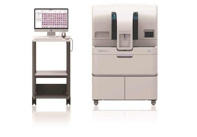
The Cobas m 511 analyzer from Roche. Not available in the United States.
Roche, Basel, Switzerland, has entered the hematology testing market with the launch of its new Cobas m 511 integrated hematology analyzer, now available for countries accepting the CE mark.
Featuring the company’s Bloodhound technology, the analyzer addresses the challenges of hematology testing by combining the three components of the process—a digital morphology analyzer, cell counter, and classifier—into a single streamlined system that prepares, stains, and analyzes microscopy blood slides.
“With this launch, patients will benefit from a faster and more accurate diagnosis of blood diseases as diverse as anemia and leukemia,” says Roland Diggelmann, CEO of Roche Diagnostics. “We are entering a new area of innovation with Roche in hematology testing, supporting customers with integrated and efficient laboratory solutions, which deliver increased medical value.”
The analyzer aims to provide greater accuracy and consistency of results by identifying, counting, isolating, and categorizing white blood cells, red blood cells, and platelets, then presenting the digital images of all these cell types. Using the analyzer, medical technologists will be able to concentrate their time on finding and classifying abnormal cells within patient samples. Such automation and digitalization reduces the need for resource-intensive manual microscope reviews, supports clinicians in sharing challenging cases around the world, and enables the quicker delivery of results that ultimately aid in patient diagnoses.
The company’s Bloodhound technology uses only 30 µL of blood to print a monolayer onto the slide, stains with an improved method for further analysis of cell morphology, and enables classification of cells displayed on a viewing station.
The analyzer images individual cells directly. Based on these direct images, the Bloodhound technology counts, analyzes morphology, and then classifies every cell in the viewing area to provide a standard complete blood count and five-part differential and reticulocyte count. While hematologists will continue to have the option of looking at slides under their microscopes, the system provides cell-by-cell images that, in many cases, may eliminate the need for microscopic review.
For more information, visit Roche.


Please sent me the price for the integrated hematology analyser