A recent advance in microscope imaging technology at the University of Waterloo could soon make diagnosing disease more accessible and affordable.
Farnoud Kazemzadeh, PhD, and Alexander Wong, PhD, University of Waterloo.
Developed by Waterloo researchers Farnoud Kazemzadeh, PhD, and Alexander Wong, PhD, the advance has led to a new form of spectral light-fusion microscope for capturing lightfield images in full color. Full-color images are required in pathology, as they enable the microscope user to analyze the behavior and interactions of different organisms at a scale much larger than traditional microscopes.
The newly developed microscope has no lens, and uses artificial intelligence and mathematical models of light to develop 3D images at a large scale. To get the same effect using current technologies—using a machine that costs several hundred thousand dollars—a technician is required to ‘stitch together’ multiple images from traditional microscopes.
“In medicine, we know that pathology is the gold standard in helping to analyze and diagnose patients, but that standard is difficult to come by in areas that can’t afford it,” says Wong, an associate professor of engineering at Waterloo and Canada research chair in medical imaging. “This technology has the potential to make pathology labs more affordable for communities who currently don’t have access to conventional equipment.”
The current spectral light-fusion microscope represents the second generation of the technology, which Wong and Kazemzadeh patented last year.
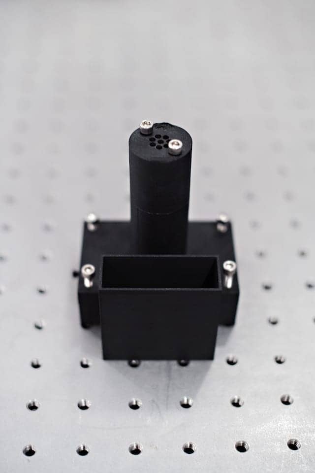
Spectral light-fusion microscope from University of Waterloo.
The microscope captures light fields and allows for 3D images that are approximately 100x larger than the 2D images captured by traditional microscopes.
“Currently, the technology required to operate a pathology lab is quite expensive and is largely restricted to places such as Europe and North America, which can afford them,” says Kazemzadeh, an adjunct professor of systems design engineering at Waterloo. “It would be interesting to see what a more affordable, mobile pathology lab could achieve.”
Details of the first-generation microscope invented by Kazemzadeh and Wong were published last year in Scientific Reports.1
REFERENCE
1. Kazemzadeh F, Wong A. Laser light-field fusion for wide-field lensfree on-chip phase contrast microscopy of nanoparticles. Sci Rep. 2016;6:38981; doi: 10.1038/srep38981.


