
Nano-Signature Discovery Could Revolutionize Cancer Diagnosis
A quick and easy test to detect cancer from blood or biopsy tissue could eventually result in a new approach to patient diagnosis. The test has been developed by University of Queensland researchers Abu Sina, PhD, Laura Carrascosa, PhD, and Matt Trau, PhD, who have discovered a unique DNA nanostructure that appears to be common to all cancers.1
Cancer is an extremely complicated and variable disease and different types of cancer have different signatures. Consequently, it has previously proven difficult to identify a simple signature that is distinct from healthy cells yet common to all cancers. The newly discovered nanostructure appears to fill the bill.
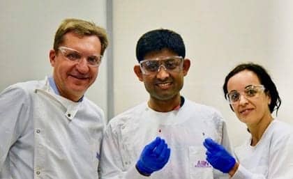
University of Queensland researchers (from left): Matt Trau, PhD; Abu Sina, PhD; and Laura Carrascosa, PhD. Image courtesy University of Queensland.
“This unique nano-scaled DNA signature appeared in every type of breast cancer we examined, and in other forms of cancer, including prostate, colorectal, and lymphoma,” says Sina. “The levels and patterns of tiny molecules called methyl groups that decorate DNA are altered dramatically by cancer. These methyl groups are key for cells to control which genes are turned on and off.”
The researchers took a holistic approach and developed a tool that could look at the nanoscale pattern changes at the whole-genome level within minutes. “In healthy cells, these methyl groups are spread out across the genome,” says Carrascosa. “But the genomes of cancer cells are essentially barren except for intense clusters of methyl groups at very specific locations.”
Gold substrate reading a normal tissue sample. Image courtesy University of Queensland.
Cancer cells release DNA fragments into blood plasma when they die. The team discovered that intense clusters of methyl groups placed in a solution caused these cancer DNA fragments to fold into unique three-dimensional nanostructures that could easily be separated by sticking to solid surfaces such as gold.
“We designed a simple test using gold nanoparticles that instantly change color to determine if the 3D nanostructures of cancer DNA are present,” says Trau. “So we were very excited about an easy way of catching these circulating free cancer DNA signatures in blood.
“Discovering that cancerous DNA molecules formed entirely different 3D nanostructures from normal circulating DNA was a breakthrough that has enabled an entirely new approach to detect cancer noninvasively in any tissue type, including blood,” adds Trau. “This led to the creation of inexpensive and portable detection devices that could eventually be used as a diagnostic tool, possibly with a mobile phone.”

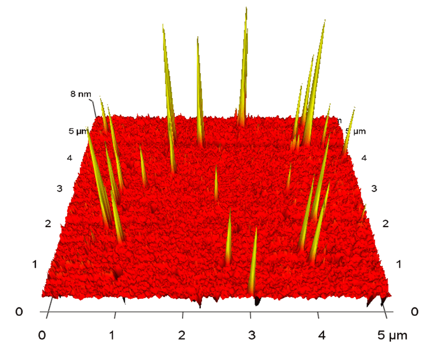
Gold substrate reading a tissue sample with cancer DNA, represented by spikes. Image courtesy University of Queensland.
The new technology has proved to be up to 90% accurate in tests involving 200 human cancer samples and normal DNA.
“We certainly don’t know yet whether it’s the holy grail for all cancer diagnostics, but it looks really interesting as an incredibly simple universal marker of cancer, and as an accessible and inexpensive technology that doesn’t require complicated lab-based equipment like DNA sequencing,” says Trau.
The researchers are working with UniQuest, the University of Queensland’s commercialization company, to further develop the technology and license with a commercial partner.
Reference
- Sina AAI, Carrascosa LG, Liang Z, et al. Epigenetically reprogrammed methylation landscape drives the DNA self-assembly and serves as a universal cancer biomarker. Nature Communications. 2018;9:4915; doi: 10.1038/s41467-018-07214-w.
Progentec Diagnostics Raises Funds to Support Advances in Lupus Detection and Management
Diagnostics innovator Progentec Diagnostics Inc, Oklahoma City, recently completed a second round of funding co-led by i2E and Chicago-based OCA Ventures. NMC Lifesciences, a leading global healthcare provider, also participated in the round.
The funding will help Progentec to commercialize its tool for identifying flare-ups of systemic lupus erythematosus (SLE) and a biomarker-based disease-activity index. Technology created by the Oklahoma Medical Research Foundation is at the core of the platform being developed by Progentec.
The Lupus Foundation of America estimates that there are as many as 1.5 million SLE patients in the United States. Seen mostly in women between the ages of 15 and 44, lupus causes the immune system to recognize and attack the body’s own tissues. Lupus sufferers have periods of flares and remission, with organs typically affected including the cardiovascular system, kidneys, lungs, reproductive organs, and skin.

Brett Adelman, Progentec.
Progentec also announced its acquisition of LupusCorner, a patient-empowerment platform for people with SLE and lupus nephritis. By integrating LupusCorner’s technology platform and data insights, Progentec is positioned to develop a first-in-class lupus management platform. The founders of LupusCorner, Arif Sorathia and Brett Adelman, have joined Progentec and will help lead technology, outreach, and growth initiatives.
Progentec’s technologies include highly accurate biomarker-based tests to diagnose the disease before symptoms begin to show, as well as tests to monitor and predict lupus disease activity levels.

Mohan Purushothaman, Progentec.
“We’ve made tremendous progress in developing a tool for the identification of lupus flare-ups before they occur,” says Mohan Purushothaman, CEO of Progentec. “Today’s funding round is the next step on our journey to making this and other advanced tools commercially available to patients with lupus, a disease that afflicts more than one million Americans, many of whom are women. The Progentec tests will become powerful tools to help patients and healthcare providers stay ahead of lupus.”
LupusCorner was founded in 2016 with the mission of transforming the lupus patient experience by creating easy-to-use and powerful technologies. Today, nearly 45,000 people with lupus interact with the LupusCorner platform each month.

Arif Sorathia, Progentec.
“LupusCorner is a powerful platform that connects people battling lupus in an online patient community to share information and better manage the disease,” says Adelman. “Our goal is to empower our users to be active participants in their healthcare, and we are excited to be part of Progentec. This is a giant step towards uniting patient-generated data with clinical measures to improve patient outcomes.”
“The addition of LupusCorner and its users furthers our vision of creating a comprehensive disease management platform and ensures that the patient voice will be a valued part of our process,” says Purushothaman. “We look forward to advancing the platform and fulfilling the unmet need for lupus diagnostics and management tools.”
For further information, visit Progentec Diagnostics.
Partners Seek to Unlock the Complexities of Cancer Biology
Melanoma specimen stained with the Bond Rx PD-L1 kit by Leica Biosystems.
Leica Biosystems, Wetzlar, Germany, has entered into a partnership to comarket UltiMapper immunohistochemistry assays by Ultivue, Cambridge, Mass, with Leica’s Bond Rx research use only staining platform.
Under the agreement, Ultivue’s UltiMapper single-step assays have been preoptimized for staining on the Bond Rx platform, creating a fully automated plug-and-play solution. The combined platform enables users to create 30 five-color, high-performance, multiplexed immunohistochemistry slides in less than 6 hours, all while preserving tissue morphology and integrity.
Ultivue’s unique DNA barcode attached to each antibody enables all antibodies to be stained in a single step, transforming a multiday manual process into an automated process that can be performed in a standard work day. UltiMapper and Bond Rx complement one another, eliminating the need for researchers to perform difficult manual operations, and instead enabling them to focus on analyzing test results.
Leica Biosystems offers researchers a complete end-to-end workflow solution. Automated staining of UltiMapper assays on the Bond Rx platform, and slide scanning and quantification using the Aperio Versa fluorescence tissue scanner and Aperio Cellular IF algorithm, brings new technologies to more researchers.

The Bond Rx research use only staining platform by Leica Biosystems.
“With tissue sample sizes decreasing, and scientific advancements expanding the number of actionable biomarkers, multiplexing is an increasingly common technique used by researchers to explore complex biology,” says Colin White, PhD, global vice president for advanced staining at Leica Biosystems. “We are very pleased to be able to offer investigators an automated version of Ultivue’s UltiMapper multiplexing technology on the Bond Rx, thereby supporting research excellence through workflow efficiency and stain consistency.”
By developing a single set of novel, proprietary reagents used for both biomarker discovery (high content, low throughput) and clinical use (lower content, high throughput), Ultivue is connecting the insights gained from research directly into the pathology lab. Applied to tissue biopsy samples, UltiMapper multiplexed assays enable simultaneous quantitation of multiple biomarkers with subcellular spatial resolution, and fit completely within traditional immunohistochemistry workflows.
Translational and clinical researchers leverage UltiMapper assays to elucidate complex biology and demonstrate their clinical utility as precision medicine tools. Ultivue is expanding its UltiMapper assay portfolio and menu of contract research services to provide a comprehensive set of personalized medicine solutions for oncology and other therapeutic areas.
“We are delighted to partner with Leica Biosystems to bring to the research market a complete, automated solution to realize the full potential of tissue-based, multiplexed marker studies at high throughput,” says Philippe Mourere, senior vice president of commercial operations at Ultivue. “Through our growing immunooncology portfolio of kits and assays, translational and clinical researchers have immediate access to unbiased biology with the ability to colocalize markers on single cells, and to functionally characterize cellular interactions within the tumor microenvironment. Looking ahead, we are expanding our level of multiplexing and widening our content offering to adjacent research areas.”
For further information, visit Leica Biosystems and Ultivue.
Featured image: Gold substrate reading a tissue sample with cancer DNA, represented by spikes. Image courtesy University of Queensland.

